Before & After Photos
Review photos of cases and see our
 most recent work.
Book a Visit
Close

Request a Consultation

Thank you!

Your submission has been received!
We will be in touch soon.
Oops! Something went wrong while submitting the form.

Request a Consultation

Thank you!

Your submission has been received!
We will be in touch soon.
Oops! Something went wrong while submitting the form.

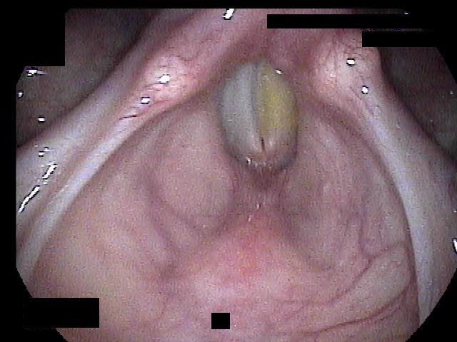
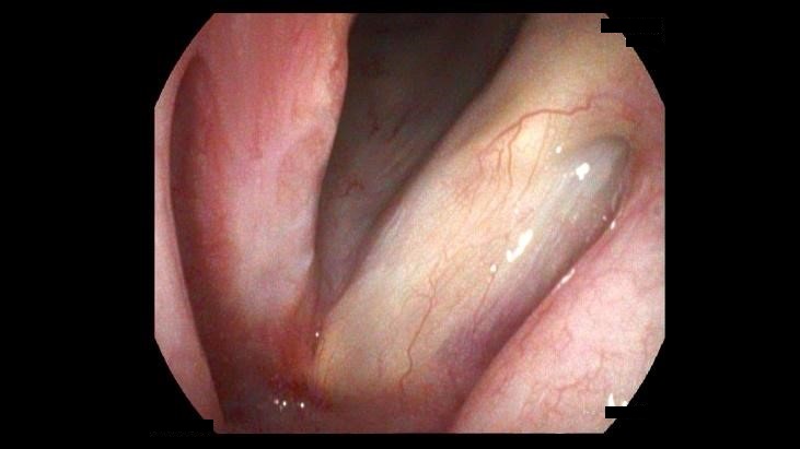
Vocal Fold Polyp

Vocal fold polyps are considered phonotraumatic lesions which are lesions that form from voice abuse, misuse or overuse. Often the inciting factor is hemorrhage from a micro vessel into the lamina propria that stretches the epithelium of the vocal fold forming the finger-like outpouching. The blood dissipates over time, but the outpouching may remain requiring surgical intervention and often voice therapy to correct the traumatic voice habits. This is a case of a middle school teacher who had persistent voice changes for several years

Before

After

Before

After

Reinke’s

Reinke’s is smoke-related polypoid degeneration of the vocal fold due to chronic smoke exposure. This is a build-up of gelatinous material in Reinke’s space a space in the subepithelial compartment of the vocal fold. This results in a deep gravelly voice change mainly affecting female smokers. In general, the mere act of quitting smoking is enough to “shrink” these changes. However, in some, the change may be permanent or critically narrows the patient’s airway. This is a case of a female smoker in her 40’s who was struggling to breathe due to these changes and thus surgery was recommended. She subsequently quit smoking and remains smoke-free.

Vocal Process Granuloma

Vocal process granulomas are traumatic lesions that usually require a combination of factors to form. The most common are excessive or repetitive coughing, prolonged intubation, traumatic intubation, repetitive shouting or screaming. Conditions like laryngopharyngeal reflux or vomiting, smoking and diabetes make these more likely to form. Often eliminating or modifying behavior (strict low-acid diet) allows these to resolve. However, in some cases these can progress. This case is a young female in her 30’s with diabetes who was intubated due to COVID-19. She presented 6 months later with already partial loss of motion to her left vocal fold suggesting progression. Thus, surgery was performed.

Before

After

Before

After

Vocal Fold Cysts

Vocal fold cysts are mucous/fluid-filled sacs that develop on the vocal folds, often as a result of vocal strain, misuse, or trauma. These cysts can vary in size and may appear on one or both vocal folds, potentially leading to symptoms such as hoarseness, breathiness, or a reduction in vocal range. While they can occur in individuals of any age, they are more commonly seen in professional voice users. Successful surgical treatment involves removing the entire sac completely without puncturing it during surgery. As a result, often a small scar may be seen at the site of excision.

VENTRICULAR CYSTS

Ventricular cysts or saccular cysts are a benign lesions that develop in the laryngeal ventricle, which is the space between the true vocal folds and the false vocal folds. These mucous filled cysts can arise from ductal obstruction of the minor salivary glands or as a result of chronic irritation or vocal overuse. They can be seen in smokers, wind instrument players and glass blowers (any activity that increases pressure and irritation in the larynx). They are similar to, but different from laryngocoeles which are filled with air and communicate with the laryngeal cavity.

Before

After

Before

After

SUPRAGLOTTIC STENOSIS

Supraglottic stenosis is narrowing of the airway at the level ABOVE the vocal folds. This usually arises from either scarring from some other procedure, radiation, or due to autoimmune conditions such as sarcoidosis or pemphigoid. This case was a 20-year-old female who reported chronic breathing difficulty. Surgical removal of the obstructing tissue was sent for pathology and this is how the patient’s sarcoidosis was diagnosed.

GLOTTIC STENOSIS

Glottic stenosis is narrowing of the airway at the level of the vocal folds. Often this results when bilateral vocal process granulomas progress and form a bridge adjoining the back of both vocal folds. This bridge then tightens slowly with time, until the patient’s airway becomes critically narrowed. Treatment is geared towards airway stabilization often at the expense of a good voice since voice and airway work in opposite directions.

Before

After

Before

After

POSTERIOR GLOTTIC STENOSIS

These images show the “scar band” that slowly tightens an airway if it is not identified and treated. This patient initially had bilateral vocal process granulomas that progressed rapidly to the scar bridge shown. These images also highlight why it is important for Dr. Syamal to get a very close look with the camera in the office as these scar bridges can be missed if the camera does not go far enough down the airway.

INFRAGLOTTIC STENOSIS

Infraglottic stenosis is a special type of airway stenosis where the narrowing begins at the inferior (undersurface) of the vocal folds. It is most seen from prolonged intubation or in the autoimmune condition called Granulomatosis with Polyangiitis (GPA, formerly known as Wegener’s). This is a case where a highly active marathon runner in her early 40’s reported increasing difficulty breathing while running. Simple reduction of just one side of her stenosis brought her back to nearly baseline.

Before

After

Before

After

SUBGLOTTIC STENOSIS

Subglottic stenosis is narrowing of the airway at levels BELOW the vocal folds. This is the single most important reason why Dr Syamal’s first scope in the office “dunks” the scope into the windpipe for a few seconds. Stenosis in this region may be due to autoimmune conditions, or from previous trauma/intubations, or idiopathic (i.e. unknown causes). This is a case of a female in her mid-40’s who had a chronic cough that was present for over 8 months. A CT scan missed the stenosis because the cuts were not fine enough and she had seen several specialists before coming to us. Exam in the office with tracheoscopy revealed the stenosis and the patient underwent surgery, and her airway remained patent thereafter.

IMPLANT MEDIALIZATION FOR VOCAL FOLD PARALYLSIS

Several options exist if a patient is found to have a vocal fold paralysis on laryngeal exam. A silastic implant is the best long-term option for a patient especially if the vocal fold closure gap is large. Dr Syamal is one of only a few voice surgeons in the United States that entirely hand-carves voice implants for each and every patient. She utilizes information from your exam, imaging and trigonometric equations to arrive at a completely unique implant size and shape designed just for you! This guarantees the best chance of matching a pre-paralysis voice. This patient was a young singer who had a paralyzed vocal fold from a nerve tumor operation when she was a child as a result her vocal folds could barely touch/close prior to surgery. Note that her vocal folds touch or “close” after the surgery.

FAT INJECTION MEDIALIZATION FOR VOCAL FOLD PARALYLSIS

Another option for a patient with vocal fold paralysis is fat injection. This is best when the vocal fold closure gap is small. This may also be used in cases of vocal fold atrophy where motion is still intact. Fat is harvested from the abdomen in and injected into the vocal fold.

HYALURONIC ACID INJECTION FOR VOCAL FOLD PARALYSIS

When a vocal fold paralysis is new, there is a small chance function may be regained. As such, we usually offer a temporary injection of a filler (hyaluronic acid) to improve the voice while we wait to see if the motion will return. In experienced hands, studies show that hyaluronic acid can last anywhere from 9-12 months. Dr Syamal has had patients where it has lasted even longer.

Source: Bertroche JT, Radder M, Kallogjeri D, Paniello RC, Bradley JP. Patient-defined duration of benefit from juvederm (hyaluronic acid) used in injection laryngoplasty. Laryngoscope. 2019 Dec;129(12):2744-2747. doi: 10.1002/lary.27842. Epub 2019 Jan 30.

Before

After

GLOTTIC CARCINOMA IN-SITU

Glottic carcinoma-in-situ is the earliest type of cancerous cells that occur on the vocal folds. This condition is characterized by the presence of abnormal cells confined to the epithelium of the vocal folds, without invasion into deeper tissues. Common symptoms include hoarseness, voice changes, and sensation changes. Glottic carcinoma in situ is often associated with risk factors such as tobacco use or hazardous exposures. Treatment is staged surgical resection until negative.

T1 GLOTTIC CARCINOMA

A T1 glottic cancer is invasive but vocal fold motion is preserved. It can involve one or both vocal folds as well as the commissures. This case involved both vocal folds and the anterior commissure. Treatment may be surgical or in some cases radiation may be selected due to extent of disease and voice preservation concerns

Before

After

Before

After

T2 GLOTTIC CARCINOMA

A T2 glottic carcinoma extends to the supraglottis or subglottis and/or affects vocal fold motion. Treatment may be surgical or radiation.

AMYLOIDOSIS

Laryngeal amyloidosis is a rare condition characterized by the accumulation of amyloid protein deposits in the tissues of the larynx, which can lead to structural changes and disruption of normal function. While laryngeal amyloidosis can occur as a localized disease, it may also be a part of systemic amyloidosis, affecting multiple organs. Diagnosis typically involves a combination of laryngoscopy and biopsy to confirm the presence of amyloid deposits. Treatment aims to alleviate symptoms and may include surgical removal of affected tissues or, in some cases, systemic therapy to address underlying amyloidosis. This patient came to us after another surgeon had already operated on the patient, but the lesion had appeared to grow back. Our surgical intervention led to her diagnosis of amyloidosis. The patient was then referred to an academic center that specialized in treatment of systemic disease.

Before

After

INFLAMMATORY MYOBLASTIC TUMOR

Inflammatory myofibroblastic tumor (IMT) is a rare tumor composed of mesenchymal myofibroblastic spindle cells enveloped by an inflammatory infiltrate. IMT is considered a borderline neoplasm with uncertain malignant potential. Treatment is complete surgical excision with monitoring for recurrence. This was a young 22-year-old female reporting a persistent cough after COVID-19. Surgical removal was successful.

LARYNGEAL PAPILLOMA

Laryngeal papilloma is caused by infection with human papillomavirus (HPV), particularly types 6 and 11. These growths can manifest as warty lesions and can occur in both children and adults, with the juvenile form often presenting more aggressively. Symptoms may include hoarseness, a raspy voice, difficulty breathing, and a sensation of a lump in the throat. Depending on their size and location, laryngeal papillomas can obstruct the airway and require treatment. Management often involves surgical removal, which may need to be repeated due to the tendency for recurrence. Regular monitoring is essential to manage symptoms and address any growth of the papillomas. While generally non-cancerous, the presence of these lesions necessitates ongoing evaluation, particularly in high-risk populations.

Before

After

Before

After

LARYNGEAL SCHWANNOMA

Laryngeal schwannoma is a rare, benign nerve sheath cell tumor derived from Schwann cells. These tumors can occur in various parts of the larynx and may lead to symptoms such as hoarseness, voice changes, difficulty breathing, or a sensation of a lump in the throat. This was a 90-year old patient that was reporting extreme difficulty swallowing with occasional breathing obstruction who opted for surgical removal. Her symptoms resolved following the surgery.

TRACHEAL GLOMUS TUMOR

A glomus tumor or paragangliomas are rare, neuroendocrine tumors that can arise from various structures such as nerves, veins or arteries. This case was a 78-year-old male that presented with a sensation hat there was something in his throat. This is another example of why tracheal examination is key as it revealed the friable red mass in the patient’s trachea. The patient was scheduled for biopsy but it was aborted when Dr Syamal noted that the mass was pulsating. Here is a great example of knowing when to NOT operate. The patient was referred to a larger academic center for further work-up, and embolization.

Before

After

Before

After

GRANULAR CELL TUMOR

Granular cell tumors are rare in the larynx that have pseudo-epitheliomatous hyperplasia on histopathology. More than 98% of these tumors are benign, but 1% to 2% of all cases occur as malignant tumors. This was a young patient who had surgery at another institution where pathology revealed a granular cell tumor. She had no further follow-up there and she presented to us a year later with life-threatening shortness of breath. She underwent immediate surgical removal with 2 additional surgeries to clear all margins of tumor.